- MENU
- HOME
- SEARCH
- WORLD
- MAIN
- AFRICA
- ASIA
- BALKANS
- EUROPE
- LATIN AMERICA
- MIDDLE EAST
- United Kingdom
- United States
- Argentina
- Australia
- Austria
- Benelux
- Brazil
- Canada
- China
- France
- Germany
- Greece
- Hungary
- India
- Indonesia
- Ireland
- Israel
- Italy
- Japan
- Korea
- Mexico
- New Zealand
- Pakistan
- Philippines
- Poland
- Russia
- South Africa
- Spain
- Taiwan
- Turkey
- USA
- BUSINESS
- WEALTH
- STOCKS
- TECH
- HEALTH
- LIFESTYLE
- ENTERTAINMENT
- SPORTS
- RSS
- iHaveNet.com: Health
Harvard Women's Health Watch
Although mammography remains standard for breast cancer screening, several newer technologies are helping to fine-tune diagnosis.
Doctors have been making radiologic images of women's breasts for almost a century. The first, taken in 1913, were x-rays of breasts that had been surgically removed. In studying these early images, doctors found that they could distinguish malignant from benign tissue, even when cancer hadn't been suspected. Here was a possible alternative to the then-standard detection method -- removing a palpable lump (if not the entire breast) and examining the tissue under a microscope.
At first, breast X-rays were disappointing. The images were often blurred by the beating of the heart, and it was difficult to distinguish tumors from breast ducts. But decades of research eventually produced x-ray equipment specifically designed for breast imaging. The new machines used low-energy x-rays and produced crisper images. New devices compressed the breast between two plates, reducing breast movement and smoothing out the tissue. By the late 1960s, mammography as we know it had been born.
Since then, new imaging methods have been developed, and today, a variety of technologies -- including ultrasound, magnetic resonance imaging (MRI), and molecular imaging -- are used to examine the breast.
MAMMOGRAPHY
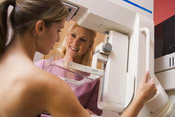
Mammography remains the "gold standard" screening method for women at average risk for breast cancer. It's relatively inexpensive, requires only a low dose of radiation, and reliably identifies malignant tumors, especially those that are too small to feel. It can also be used to investigate breast lumps and other symptoms.
On regular mammograms, fat looks dark gray, and breast tissue, which is denser, is white. Abnormalities, such as lumps, also appear white, making it difficult to distinguish them from the surrounding tissue. In the digital mammogram, a cancerous mass can be seen as solid white, just behind the nipple. The tumor is harder to spot on a standard mammogram of the same breast.
How it works: X-ray radiation passes through the breast, producing an image on film or on a digital recording plate.
What it involves: Whether the mammography is film or digital, your experience will be the same. You'll remove your clothes above the waist and don an open-front hospital gown. A technologist will adjust the imaging platform to your height, arrange your breast on it, and position your arms and torso. A specially designed clear paddle will be lowered to compress your breast. For a routine screening mammogram, two images of each breast are taken -- one from above and one obliquely from the side. Diagnostic mammograms may require more images. The procedure takes anywhere from 15 minutes to an hour.
ULTRASOUND
Ultrasound imaging -- also called sonography -- may be used to evaluate abnormalities detected during a breast exam or mammogram. Though it can't detect microcalcifications (tiny flecks of calcium that may signal early cancers), it excels in distinguishing solids from liquids so it's useful for differentiating solid tumors from fluid-filled cysts, which are benign. Ultrasound can also be used to guide needle biopsies.
How it works: Ultrasound creates an image from reflected high-frequency sound waves emitted by a device called a transducer. The technician will run the transducer across the surface of the breast to produce a real-time image of its interior structures. Doppler ultrasound, which tracks the speed of blood coursing through the vessels, is also occasionally added to assess blood flow to breast lesions.
What it involves: With your clothing removed from the waist up and wearing an open-front hospital gown, you'll lie on your back on a padded table under dim light (so that the technician can get a good view of the monitor). A gel will be applied to your breast and the transducer will be placed over it. The ultrasound waves are too high a frequency to be heard, and the procedure itself is painless -- all you may feel is the slight pressure of the transducer sliding over your breast. The test takes three to 15 minutes.
Film-screen or digital mammography?
In 2000, the U.
According to the Digital Mammography Imaging Screening Trial (involving almost 50,000 women), the difference between film and digital mammography is negligible for most women over age 50, but digital images have an edge over film for three other, often overlapping groups -- women under age 50, women who are pre- or perimenopausal, and women who have dense breasts. If any of those descriptions fit you, you should talk to your clinician about the possibility of having a digital mammogram at your next screening.
Magnetic resonance imaging (MRI)
MRI using a special receiver and injected contrast material to image only the breasts is very good at detecting invasive breast cancer. However, it can also misidentify benign lesions as malignant, because both can absorb the contrast.
MRI is not a substitute for regular mammography, but the
Once a malignancy is detected, MRI may also be used to find or rule out additional tumors and thus may be helpful in deciding whether breast-conserving surgery or mastectomy is the best treatment option. Also, MRI without the use of contrast material may be employed to determine whether silicone breast implants have ruptured.
How it works: MRI uses a powerful magnetic field and radio frequency pulses that are processed by a computer to create images of organs and tissues. It does not use ionizing radiation (x-rays). The resulting digital images are examined on a computer monitor, and like digital mammograms, MRI images can be stored and transmitted electronically.
What it involves: You will change into a hospital gown and remove any metal-containing jewelry or devices (like hearing aids) that might interfere with the magnet. An intravenous (IV) catheter will be inserted into your arm or hand, and you will receive an injection of a compound containing gadolinium to outline the structures of the breast on the MRI image. Then you will lie on your stomach on a padded table, and your breasts will be positioned into padded openings within a special coil, or receiver, that detects the magnetic signal. The table is then moved into the machine that contains the magnet. You will be given earplugs or possibly headphones for listening to music during the procedure to muffle the knocking sound emitted each time an image is taken. The MRI session takes 30 to 45 minutes.
Positron emission mammography (PEM)
PEM is used in addition to mammography to identify small invasive cancers and ductal carcinoma in situ (DCIS) -- cancer that is confined to the milk ducts. PEM is not yet widely available and may not be covered by insurance.
How it works: A radioactive glucose tracer emits gamma rays that are detected by a camera. Because glucose is absorbed and stored more readily in rapidly growing cells -- like cancer cells -- a tumor accumulates higher concentrations of the tracer than healthy tissues and shows up as a "hot spot" on the photographic image. A computer analyzes the image to determine the size, shape, and location of the mass.
What it involves: You'll need to avoid eating or drinking anything except water in the six hours before the test. The technologist will check your blood glucose level with a finger-prick test, and if the results are normal, a radioactive glucose tracer will be injected into your arm. You'll wait for an hour as your body absorbs the tracer. From then on, PEM is much like a mammogram, except that you'll be seated in a chair at the scanner. Two scans are made of each breast, and the entire procedure takes about two hours.
Thermography: Not a screening option
Thermography, a technique originally designed for night-vision devices used by the U.S. military, records the temperature of the body by measuring the infrared radiation it emits. Malignant tissue generally has a higher temperature than normal tissues because of its richer blood supply and higher metabolic rate, so scientists reasoned that infrared "hot spots" in the breast might signal the presence of cancer. Thermography is approved as an adjunctive tool for diagnosing breast cancer, but it produces too many false-positive results to be useful as a screening tool.
Breast-specific gamma imaging (BSGI)
Like PEM, BSGI is used as an adjunct to mammography, is not widely available, and may not be covered by insurance.
How it works: BSGI also employs a radioactive tracer to identify cancer cells. In this case, the substance used is technetium sestamibi, a compound that accumulates in mitochondria, the power plants of the cells. Rapidly proliferating cells are rich in mitochondria, and the tracer will concentrate there, emitting gamma rays that produce dark spots on a digital image.
What it involves: Before the exam, an IV catheter will be inserted into your arm or hand. The tracer will be injected into the IV solution and you'll sit quietly for 10 minutes while it's being absorbed. The imaging process is similar to PEM. Two scans are made of each breast, and additional scans are sometimes taken of your underarm lymph nodes. The whole procedure takes about an hour.
THE FUTURE OF BREAST IMAGING
According to the
Available at Amazon.com:
Copyright © Harvard Health Letters. All rights reserved.
AGING | ALTERNATIVE | AILMENTS | DRUGS | FITNESS | GENETICS | CHILDREN'S | MEN'S | WOMEN'S
Health - Advances in Breast Cancer Screening Helping to Fine-Tune Diagnosis