- MENU
- HOME
- SEARCH
- WORLD
- MAIN
- AFRICA
- ASIA
- BALKANS
- EUROPE
- LATIN AMERICA
- MIDDLE EAST
- United Kingdom
- United States
- Argentina
- Australia
- Austria
- Benelux
- Brazil
- Canada
- China
- France
- Germany
- Greece
- Hungary
- India
- Indonesia
- Ireland
- Israel
- Italy
- Japan
- Korea
- Mexico
- New Zealand
- Pakistan
- Philippines
- Poland
- Russia
- South Africa
- Spain
- Taiwan
- Turkey
- USA
- BUSINESS
- WEALTH
- STOCKS
- TECH
- HEALTH
- LIFESTYLE
- ENTERTAINMENT
- SPORTS
- RSS
- iHaveNet.com
Claire Ainsworth, New Scientist Magazine
It starts with a barely perceptible blurring of vision from time to time -- the sort of thing you might chalk up to getting older. But when you get it checked out, there's disturbing news: You have a disease called age-related macular degeneration, or AMD.
AMD can progress slowly or quickly, but there's no cure. Your hopes for an idyllic retirement -- reading all those books, driving to new places, or just enjoying a carefree independence -- are now clouded by uncertainty.
It's a depressing picture, and the odds are that one day it will happen to you or someone you know. People typically develop AMD after the age of 50, and it affects nearly 1 in 10 people over the age of 80. It's the most common cause of blindness in the West.
That picture may be about to change, however. A decade ago, an important insight into the biology of AMD led to an explosion of new strategies to treat it. And thanks to several anatomical peculiarities of the eye, it's an ideal test bed for therapies that would be riskier in other parts of the body.
As a result, AMD has been the focus of several high-tech approaches, ranging from RNA interference to gene therapy and stem cells. Some of the most promising will soon be tested in people and could become established therapies in the next decade.
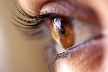
As its name suggests, AMD is a disease of the macula, a pea-sized patch in the center of the retina. Its high acuity allows you to see fine detail, such as the letters on a page.
The retina has several layers. Incoming light first reaches the photoreceptor cells, which turn it into electrical impulses. Underneath is a layer of supporting cells called the retinal pigmented epithelium, or RPE, which nourish the photoreceptors and clean up their waste. Below the RPE are the capillaries that form the retina's blood supply, but between blood and RPE is a thin layer called the blood-retinal barrier. Like the blood-brain barrier, this is only selectively permeable, keeping any toxins in the blood from reaching the delicate photoreceptors. It also helps to keep out the immune system and means that anything put into the eye is more likely to stay there.
The exact causes of AMD are still unclear, but the RPE cells appear to wither and die first. With their support system deteriorating, the photoreceptors die, too. People retain their fuzzier peripheral vision, but as the disease progresses, their central vision goes and with it the ability to read, drive or even recognize facial expressions.
"That is very isolating for people," says Barbara McLaughlan of the
For about 1 in 10 people with AMD, though, the disease suddenly progresses with terrifying speed. New blood vessels sprout under the retina, leaking blood and fluid. The macula swells and becomes scarred, and central vision can be lost in weeks. This form is known as wet AMD, while the slower version is the dry form.
Until recently, people with AMD had few options. They are advised to avoid smoking and eat a diet rich in zinc, beta-carotene and vitamins C and E, but that can only slow things down. Various forms of eye surgery and laser or light therapies are used to treat wet AMD but these are either risky or of limited help.
In the 1990s, however, studies of eye tissue taken at autopsy led to a key insight. The maculas of people with wet AMD had high levels of a signaling molecule called vascular endothelial growth factor (VEGF), which triggers the growth of new blood vessels.
SIGHT RESTORED
The idea of blocking the action of this molecule has generated a host of treatment strategies. The most successful so far is an antibody fragment that binds to VEGF. Called Lucentis, it halts vision loss in about 95 percent of people with wet AMD, and restores some sight in more than a third.
"We had never seen anything like it," says Daniel Martin of the
This approach is no panacea, though. For one thing the treatment must be injected into the eye every 4 to 6 weeks, and this occasionally triggers retinal detachment or an eye infection that can lead to blindness. It's also expensive: Lucentis, which is the only antibody-based drug approved to treat wet AMD, costs
Still, now that the principle of blocking VEGF has been established, various groups are working on improved therapies. The next treatment to reach the clinic might be VEGF Trap-Eye, a protein that binds to VEGF more tightly than Lucentis as well as mopping up another molecule involved in blood vessel growth -- placental growth factor. Developer Regeneron says this might make it more potent, or require less frequent injections, perhaps every 8 weeks. The first results from large trials comparing VEGF Trap-Eye with Lucentis are due by the end of the year.
A completely different approach is to use RNA interference, a relatively new technique for switching off individual genes by delivering short stretches of RNA. So far the main hurdle has been getting RNA into the target cells, but the eye's accessibility means this is not so much of a problem. A few RNA-based drugs designed to block AMD are in the early stages of development.
The first to be tested in people was one called bevasiranib, though so far results have been poor. Other firms are looking beyond directly blocking VEGF, and are using RNA interference to target other molecules in its signaling pathway, either upstream or downstream.
Like VEGF antibodies, RNA drugs would still entail regular eye injections. What's needed is a longer-lasting approach, which might come in the form of gene therapy.
The idea of permanently inserting new genes into cells has generated great hopes in the past few decades but it's a risky path to tread. In 1999, U.S. teenager Jesse Gelsinger died after a massive immune reaction against the virus used to deliver a gene. In 2002, a different virus triggered leukemia in several children.
The eye might be a safer testing ground for gene therapies. It's enclosed by the blood-retina barrier, keeping viruses in and the immune system mostly out. Side effects should be limited to the eye, and in a worst-case scenario, the organ could be removed without endangering life. Researchers can peer inside for easy delivery of the therapy and to monitor progress, and the other, untreated eye is the perfect experimental control. There are also simple and safe ways to test the retina's functioning. Partly thanks to work in the eye, gene therapy is seeing something of a renaissance.
Proof of principle came in 2000 when a team led by Robin Ali at the
Slowly and cautiously, Ali's group and others have been trying out an AAV-based gene therapy in people with an inherited form of blindness. There has been some vision improvement in most of the people who have received it so far. After just one treatment, benefits can persist for two years, according to Ali's latest, unpublished results.
"This gave a huge boost to the field, because it showed it can be safe and effective," says Ali.
Work has now begun to find out whether gene therapy could provide a long-lasting treatment for AMD. U.S. firm
All this is a long way from becoming an established therapy, however. In the meantime, faster progress may come via lower-tech drugs, with molecules small enough to diffuse through the tissues of the eye, so they could be given as eye drops. For example, a group of drugs called kinase inhibitors block the signals involved in blood vessel growth. Some are already used as cancer treatments and several are now in clinical trials for wet AMD, where they look promising.
Yet all these approaches have a major drawback. Because they work by blocking abnormal blood vessel growth, they would be no help for the 85 percent of AMD patients who have the dry form of the disease.
Now, however, a possible treatment for dry AMD is on the horizon. Instead of tinkering with molecules or genes, the strategy is to entirely replace the problem RPE layer with a new one grown from stem cells.
CELL REPLACEMENT
In theory, stem cells offer the hope of growing new tissues to order, yet in reality, many challenges remain. At the moment human embryonic stem cells usually come from surplus IVF embryos. Tissues derived from these would be rejected unless the patient takes lifelong immunosuppressive drugs, as after an organ transplant. But again, the eye has a unique advantage. Because it's relatively shielded from the immune system, only low doses of immunosuppressants should be needed to prevent rejection.
One of the pioneering groups in this field is led by Pete Coffey at University College London. They have grown tiny sheets of RPE cells in the lab and have shown these can restore vision in rats and pigs. They hope to start the first trials in people early next year.
"It could be an immense step," says Coffey. "No one has yet gone into the clinic with a human embryonic stem cell therapy."
U.S. firm Pfizer has teamed up with Coffey, and has made RPE cells the main focus of its new regenerative medicine unit in Cambridge, UK. "We are trying to make the production of RPE more commercial," says Ruth Mackernon, chief scientific officer at the Cambridge unit.
U.S. company ACT is also developing stem-cell RPE transplants, which have been successfully tested in animals. The company is now in talks with the
RPE transplants would only be helpful for people who still have some photoreceptors left to salvage, but one day even people beyond this stage may benefit. In 2006, Ali's team announced that they had taken photoreceptor cells from a young mouse and transplanted them into an adult mouse retina (Nature). The team is now working on doing the same with human embryonic stem cells.
It is fair to say that in the next few years the eye will see a host of pioneering therapies. Hopefully they will mean more of us can look forward to that idyllic retirement after all.
Macular Degeneration
Macular degeneration is a progressive eye condition that affects the central part of the retina, known as the macula, which is responsible for sharp, detailed vision. There are two main types of macular degeneration: dry and wet. Dry macular degeneration, which is more common, is characterized by the gradual loss of photoreceptor cells in the macula. Wet macular degeneration occurs when abnormal blood vessels grow under the macula and leak fluid, causing damage to the photoreceptor cells. Common symptoms of macular degeneration include blurred or hazy central vision, difficulty reading or recognizing faces, and changes in the appearance of straight lines. There is no cure for macular degeneration, but early detection and treatment can help slow its progression and preserve remaining vision. Treatments include anti-VEGF injections, photodynamic therapy, and vitamins and supplements.
WORLD | AFRICA | ASIA | EUROPE | LATIN AMERICA | MIDDLE EAST | UNITED STATES | ECONOMICS | EDUCATION | ENVIRONMENT | FOREIGN POLICY | POLITICS
Health - Saving Sight From Macular Degeneration: New Therapies Show Promise